INTRODUCTION
Different breast cancer subtypes, determined on the basis of gene expression patterns derived from microarray analysis, are associated with significantly different survival and recurrence rates [1,2]. However, because of the cost and time constraints, gene expression cannot be evaluated in every breast cancer patient. Therefore, a simplified classification based on immunohistochemical (IHC) analysis of estrogen receptor (ER), progesterone receptor (PR), and human epidermal growth factor receptor 2 (HER2) has been adopted in clinical practice [3]. Recently, the importance of proliferation indicators such as Ki-67 has been emphasized because several studies revealed that the Ki-67 level was associated with treatment response and survival after neoadjuvant treatment [4,5]. According to the St. Gallen International Expert Consensus on the primary therapy of early breast cancer, microarray molecular subgroup classification based on IHC analysis of ER, PR, HER2, and Ki-67 provides powerful prognostic information [3,6]. Although surgical specimens collected after breast cancer surgery provide the most reliable results for IHC evaluation of ER, PR, HER2, and Ki-67, IHC analysis of core needle biopsy (CNB) specimens is required in certain cases.
When patients with locally advanced breast cancer are considered for neoadjuvant treatment, therapeutic strategies are based on the results of IHC analysis of CNB specimens. In addition, when distant metastasis occurs after primary breast cancer treatment, it is crucial to assess the ER, PR, HER2, and Ki-67 statuses and histologically evaluate the metastatic lesions because the ER, PR, and HER2 statuses of 14% to 42% of recurrent and distant metastatic lesions are different from that of the corresponding primary breast cancer [7,8]. Although the accuracy of CNB for breast cancer diagnosis has been reported to be similar to that of open surgical biopsy, some factors including small sample size, tumor heterogeneity, and technical errors could influence the diagnostic accuracy of CNB. Previous studies have reported discordance between ER, PR, or HER2 evaluations by CNB and those by surgical biopsy [9,10].
The aim of this study was to compare the ER, PR, HER2, and Ki-67 statuses determined by CNB with those determined by subsequent surgical biopsy in order to measure the level of concordance between the results of CNBs and surgical biopsies performed at Chung-Ang University Hospital.
METHODS
Patients
We reviewed the medical records of patients who underwent breast cancer surgery at Chung-Ang University Hospital between March 2012 and December 2015 and included women with a diagnosis of ductal carcinoma in situ (DCIS) or invasive breast cancer. The inclusion criteria were as follows: (1) newly diagnosed DCIS or invasive breast cancer; (2) available pathologic data including ER, PR, HER2, and Ki-67 status determined by using both CNB and surgical specimens; and (3) no neoadjuvant chemotherapy. In 46 patients whose IHC results were missing, additional IHC analysis was performed to generate a complete ER, PR, HER2, and Ki-67 profile, after obtaining informed consent. A total of 191 patients met the inclusion criteria. This study was approved by the Institutional Review Board of the Chung-Ang University Hospital (IRB number: C20140751 [1271]).
Pathological assessment
CNB specimens were retrieved from tumor centers by using an ultrasonography-guided 14-gauge Tru-Cut needle. All immunostaining procedures and subsequent interpretations were performed at our institution. Specimen slides of patients who had undergone CNB at other clinics were reviewed again at our institution. In case of missing IHC results, additional staining and review processes were performed. The pathology results were interpreted by a senior pathologist with abundant experience in evaluating breast cancer biopsies.
IHC analysis was performed to evaluate the expression of ER, PR, HER2, and Ki-67 in CNB and surgical specimens. ER and PR expression was calculated as a percentage of cells showing definite nuclear staining for ER and PR. The cutoff value for ER and PR positivity was ≥1% of tumor cells positive for nuclear staining. HER2 expression was assessed on a scale from 0 to 3+ according to the intensity of cell membrane staining. Specimens were considered HER2-positive either when the IHC score was 3+ or when HER2 gene amplification was identified by fluorescence in situ hybridization (FISH) [11]. Ki-67 expression was measured using a rabbit monoclonal antibody as previously described, and expressed as a percentage of immunohistochemically stained cells relative to the total number of counted cells [12]. The cutoff value for Ki-67 positivity was ≥15% of the cells positive for staining.
Statistical analysis
Patient characteristics are presented descriptively as percentages. Pearson correlation coefficient was used to assess the correlation between ER, PR, HER2, and Ki-67 expression in CNB specimens and that in surgical specimens. According to the cutoff value of each biomarker, the sensitivity, specificity, negative predictive value (NPV)/positive predictive value (PPV), and false negative/positive rates of CNB were calculated, and compared with the results from surgical specimens. A two-sided t-test was used for between-group comparisons with p<0.05 being considered statistically significant. All statistical analyses were performed using SPSS version 17.0 software (SPSS Inc., Chicago, USA) and R version 3.2.3 (R Core Team, Vienna, Austria).
RESULTS
A total of 191 patients were included in this study. The patient characteristics are summarized in Table 1. According to American Joint Committee on Cancer staging seventh edition, 27 (14.1%), 101 (52.9%), 58 (30.4%), and five patients (1.6%) had Tis, T1, T2, and T3 stage breast tumors, respectively. The stage of nodal metastasis stage in 125 (65.4%), 42 (22.0%), 12 (6.3%), and seven patients (3.7%) was N0, N1, N2, and N3, respectively. In terms of histologic classification, invasive ductal carcinoma (134 patients, 70.1%) was the most common, followed by DCIS (34 patients, 17.8%) and mucinous carcinoma (12 patients, 6.3%). Among the 191 patients, 156 (81.7%) underwent breast-conserving surgery and 35 (18.3%) underwent mastectomy. In terms of axillary lymph node dissection, 138 patients (72.3%) underwent sentinel lymph node biopsy only and 48 patients (25.1%) underwent axillary lymph node dissection. Five patients did not undergo axillary node surgery (Table 1).
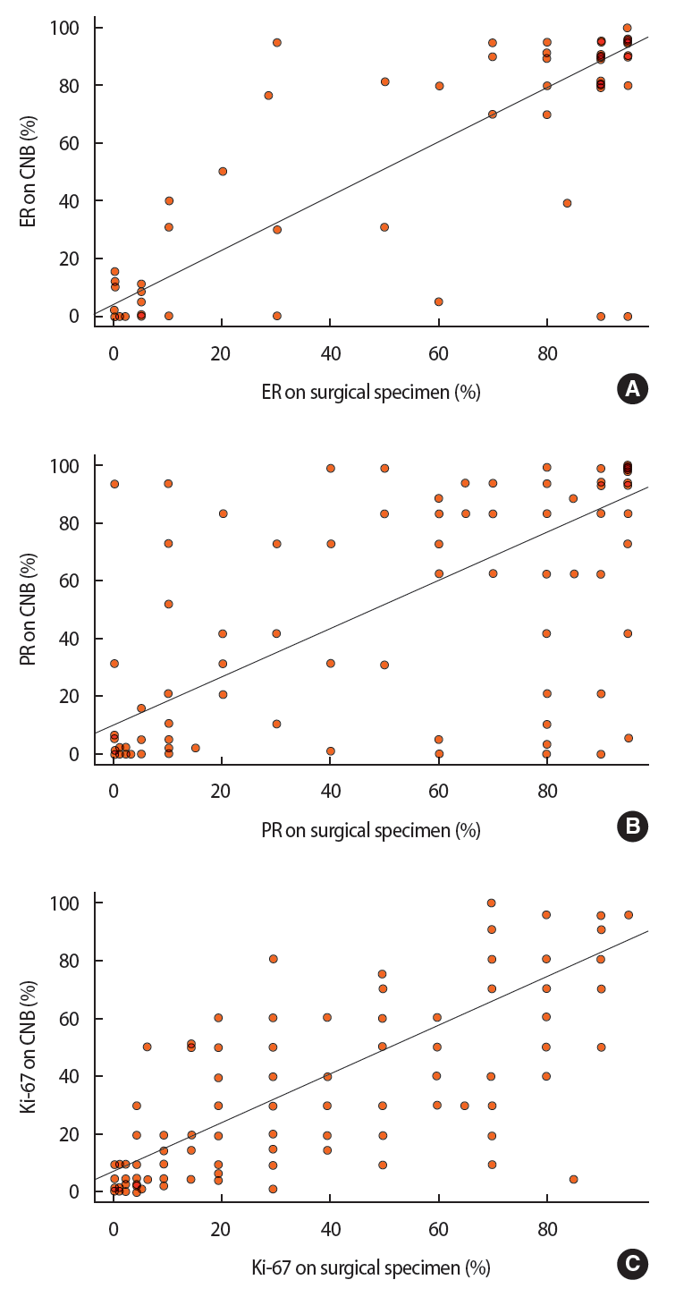
In the correlation analysis, Pearson correlation coefficients were 0.950 for ER expression (95% confidence interval [CI], 0.934–0.962; p< 0.0001), 0.813 for PR expression (95% CI, 0.759–0.856; p< 0.0001), 0.847 for HER2 grade (95% CI, 0.802–0.884; p< 0.0001), and 0.817 for Ki-67 level (95% CI, 0.75–0.857; p< 0.0001) (Figure 1).
The ER, PR, HER2, and Ki-67 statuses, based on the cutoff value of each factor, in CNB and surgical specimens are shown in Table 2. The ER status was assessed as positive in CNB specimens from 141 patients (73.8%) and surgical specimens from 148 patients (77.5%). The sensitivity and specificity of ER assessment were 92.6% and 90.7%, respectively, in CNB specimens. The PPV and NPV of ER status were 97.2% and 78.0%, respectively, in CNB specimens. PR status was assessed as positive in CNB specimens from 127 patients (66.5%) and surgical specimens from 134 patients (70.2%). The sensitivity and specificity of PR assessment were 88.8% and 86.0%, respectively, in CNB specimens. The PPV and NPV of PR were 93.7% and 76.6%, respectively, in CNB specimens. Ki-67 expression was assessed in CNB and surgical specimens from 179 patients. The Ki-67 level was not available in 12 patients because of the lack of availability of samples. Ki-67 expression was positive in CNB and surgical specimens from 95 patients (53.1%) and 108 patients (60.3%), respectively. The sensitivity and specificity of Ki-67 assessment were 80.6% and 88.7%, respectively, in CNB specimens. The PPV and NPV of Ki-67 were 91.6% and 75.0%, respectively, in CNB specimens.
HER2 status was assessed in 152 patients because samples were not available and HER2 assessment was unnecessary in DCIS or very small tumors in invasive breast cancer. Among 23 patients whose HER2 was 2+ on IHC, four showed HER2 gene amplification in FISH and were thereby categorized as HER2-positive. CNB specimens from 37 patients (24.3%) and surgical specimens from 36 patients (23.7%) were HER2-positive. There was only one discordant case, which was positive in the CNB specimen but negative in the corresponding surgical specimen. The sensitivity and specificity of HER2 in CNB specimens were 100% and 99.1%, respectively. The PPV and NPV of HER2 in CNB specimens were 97.3% and 100%, respectively (Table 3).
DISCUSSION
CNB is a standard diagnostic procedure conducted in patients with suspected breast cancer. Although the histopathological diagnosis of breast cancer from CNB specimens is of prime importance, IHC evaluation of biomarkers such as ER, PR, HER2, and Ki-67 offers additional information that helps determine the optimal therapeutic regimen. Our results showed high concordance rates between the results from CNB and surgical specimens: 92.2%, 88.0%, 99.3%, and 83.8% for ER, PR, HER2, and Ki-67 expression, respectively. The ER positivity rate was higher in CNB specimens than in surgical specimens, which is consistent with the results of a previous study [9].
The concordance rate of ER expression was slightly higher than that of PR, which was consistent with previous reports [10,13,14]. The distribution of PR in tumors is more heterogeneous than that of ER [9,15]. Studies using both IHC and FISH to evaluate HER2 status had higher concordance rates than those using IHC alone [10,16,17]. We tried to perform additional FISH tests in 2+ cases and evaluate HER2 amplification. However, because of time and cost constraints, most patients with HER2 2+ results did not undergo FISH tests. In our study population, the positivity rate of HER2 was 24.3% in CNB specimens and 23.7% in surgical specimens, which was slightly higher than the rates reported in previous studies [14].
In this study, the concordance rate for Ki-67 was lower than that for other biomarkers, indicating that Ki-67 is more heterogeneously distributed [14]. The concordance rates of Ki-67 were similar when the Ki-67 expression cutoff values were 14% and 20% (82.4% and 81.3%, respectively). There was an increase in Ki-67 expression after CNB. Tagliabue et al. [18] have suggested that the post-CNB increase in the expression of Ki-67, a well-known predictive marker for tumor proliferation, might be related to the wound healing process. An increase in Ki-67 expression has been reported in specific breast cancer molecular subtypes: HER2-positive and triple negative tumors [18,19].
More than 30 single-center studies have compared the predictive validity of percutaneous diagnostic biopsies to that of surgical specimens in determining histologic type and other biomarkers [9,10,13-17,20,21]. Although the concordance rates did vary among these studies, their results consistently indicated that preoperative determination with CNB has a high concordance rate. Some of these studies compared ER expression alone, whereas some included ER, PR, and HER2 [9,10,13,14,17,21,22]. Chen et al. [23] performed a meta-analysis of 27 studies and concluded that CNB has high concordance rates with excisional biopsy. The included studies had different sample sizes, sample number of biopsies, cutoff value of positivity, and technical differences in biopsy procedures and staining.
Several explanations have been proposed previously for the discordance between the results of CNB and those of surgical specimens. First, because cancer has a heterogeneous character, the core does not represent the entire tumor [22]. Factors related to intratumoral heterogeneity, such as larger tumor size, axillary lymph node involvement, younger patient age, and lower grade of tumor, have been shown to contribute to a higher discordance in the results [16,21,24]. Second, proper formalin fixation is essential for evaluating IHC status. An increase in the duration of formalin fixation is associated with an increase in the number of false-negative results and a sample fixation time of less than 24 hours is recommended [16,25]. We attempted to limit the fixation time to less than 12 hours for samples retrieved from our institution. However, any technical errors during the entire process can affect the results of IHC status.
Adjuvant chemotherapy, which is widely used in breast cancer treatment, has the advantages of lower cancer recurrence and improved overall survival [26]. Application of endocrine therapy is also increasing because it offers the benefits of safety and ease of administration [27]. In HER2-positive cancers, which are known for unfavorable prognoses, the anti-HER2/neu receptor antagonist trastuzumab improves disease-free survival and overall survival [28]. However, as trastuzumab has no benefits in HER2-negative breast cancers, a careful determination of HER2 status and selection of appropriate therapeutic agents is crucial. Severe complications such as cardiac toxicity can occur during the administration of trastuzumab, resulting in an increase in the cost of care. During preoperative chemotherapy, including hormonal therapy or targeted therapy, clinicians depend on the CNB results for the selection of optimal therapeutic agents. Performing biomarker tests on CNB specimens might not be cost effective for patients who do not receive neoadjuvant therapy. However, future studies are warranted to improve the precision of the assessment and classification of the IHC status of breast cancer in both diagnostic biopsy and surgical specimens.
Our study has some limitations. First, a sample size about 200 patients might not be sufficient for reliable statistical analysis. Second, although all breast cancer types, including precancerous DCIS lesions, were analyzed in this study, the low number of invasive ductal carcinoma cases presented some difficulties during sub-analysis. Third, the CNB-based diagnosis of some patients was performed at local clinics. Although we performed an additional review of the slides at our hospital and checked the IHC status, the staining protocols used at these clinics and our institution might have been different.
We conclude that, compared with surgical specimens, CNB has high diagnostic accuracy in evaluating ER, PR, HER2, and Ki-67 status. Our findings support the recommendation that CNB should be considered as the initial procedure for assessment of receptor status in breast cancers.