INTRODUCTION
Sparganosis is a rare parasitic infection caused by the larvae of Spirometra erinace [1]. Humans are an uncommon host species for the parasite, but transmission can occur by drinking contaminated water or by eating raw or partially cooked fishes, snakes, or frogs [2]. After humans are infected with spargana, the larvae can infect the abdominal wall, extremities, urogenital system, and/or the central nervous system [3,4]. Patients usually present with a subcutaneous mass or vague pain [1]. Sparganosis can recur depending on the location of the infection site and the incompleteness of the excision [1]. About 70% of patients are only infected with a single sparganum, therefore recurrent sparganosis is a rare disease [5]. In this study, we report a case of recurrent breast sparganosis that occurred two years after the surgical excision of worms from the ipsilateral breast. As spargana can survive in the human body for an extended period of time [6], regular follow-up of patients who were treated for sparganosis should be part of an obligatory treatment plan.
Ethical issues in this study were confirmed by the Institutional Review Board (IRB) at author’s institution (No. INHAUH 2019-04-017).
CASE REPORT
A 70-year-old woman visited a local clinic in 2016 with a complaint about a palpable mass in the upper outer quadrant of her right breast. She had first noticed the palpable breast mass in 2014, but she did not visit a clinic for an assessment. In 2016, she noticed that the size of the mass had increased and subsequently visited a local clinic. After sonographic evaluation, she was transferred to our clinic for a biopsy of the breast mass.
The patient had no prior history of breast trauma or breast cancer. Furthermore, she had no history of eating snakes or frogs. However, she did have a history of drinking unpurified water, such as water from a mineral spring. Physical examination of her right breast revealed a firm mass in the 10 o’clock position. Laboratory test results were indicative of mild eosinophilia (8.8%). There was no evidence of axillary lymphadenopathy.
Sonography performed in our clinic revealed tortuous, multi-tubular, hypoechoic structures in the right breast (Figure 1A, 1B, and 1C). The appearances of the structures were suggestive of sparganosis, and we undertook a core needle biopsy. The result of core needle biopsy revealed a parasitic worm with dense lymphoplasmacytic and eosinophilic infiltrations. We surgically removed the mass and the ivory-white, ribbon-like parasites from her right breast. Final pathology confirmed the parasite as spargana (Figure 1D).
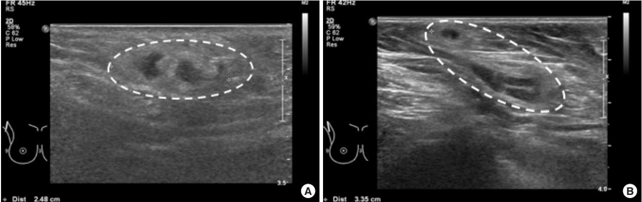
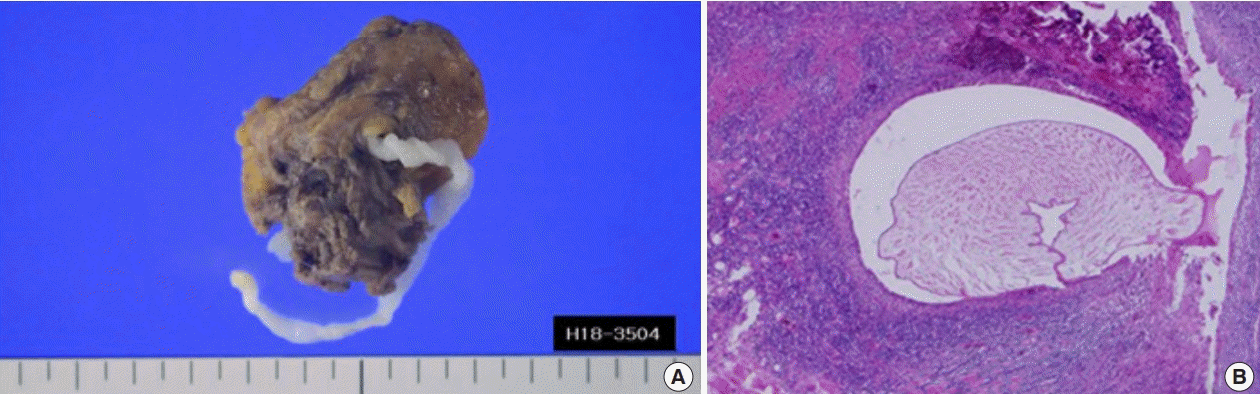
Two years later (2018), the patient again visited our clinic with a complaint of a palpable mass in her right axillary fossa. Breast sonography showed tubular structures of approximately 2.48 cm and 3.35 cm in the right superficial axillary fossa, which were determined to be indications of sparganosis (Figure 2). We surgically excised the masses from her right axilla and confirmed a diagnosis of recurrent sparganosis (Figure 3A). The pathology report determined that the patient had sparganosis with chronic granulomatous inflammation and eosinophilic infiltration (Figure 3B). We concluded that the recurrent sparganosis was a result of either incomplete removal of parasites during the previous operation or of parasite re-infection.
DISCUSSION
Sparganosis is an uncommon parasite infection in humans caused by the plerocercoid larvae of Spirometra erinacei [1]. The routes of transmission include drinking contaminated water or eating raw or partially cooked fish, snakes, or frogs [2]. Sparganum can migrate to any part of the body, including the abdominal wall, extremities, urogenital system, central nervous system, or the orbital region [3,4].
Breast sparganosis has been reported intermittently since 1981 and its incidence is less than 2% of all reported cases of sparganosis [7]. Although it is important to elucidate the mode of infection in cases of breast sparganosis, the majority of reported breast sparganum infections have unclear sources of infection [4]. In the present case, the presumed cause of infection was drinking water contaminated with the parasites.
Breast sparganosis has characteristic radiologic indicators. When radiologic studies detect multilobulated, elongated tubular structures in the subcutaneous space or retromammary space, breast sparganosis should be suspected [7]. Mammography may reveal a radio-opaque structure in a fat layer without calcification, whereas sonography can show elongated, tubular, and hypoechoic structures with or without hetero-echogenicity [1]. However, sonographically detected elongated tubular structures can be confused with duct ectasia, radiation edema, superficial thrombophlebitis, or congestive heart failure [1,7,8]. Thus, it is necessary to distinguish breast sparganosis from other diseases when sonography shows perilesional hyperechogenicity due to chronic granulomatous inflammation [8].
Currently, the majority of breast lesions undergo core needle biopsies or vacuum-assisted biopsies. However, in breast sparganosis cases, the need to perform a preoperative biopsy has been the subject of controversy. Some authors have suggested that biopsy procedures prior to surgery may lead to the fragmentation of the tapeworms, thereby increasing the incidence of recurrence due to incomplete surgical removal [7,9]. We recommend that, in patients with suspected breast sparganosis, surgical removal should be considered instead of cutaneous biopsies, as previously suggested [9].
For breast sparganosis, medical treatment with praziquantel should be included in secondary treatment plans for cases with a surgically unresectable infection, as praziquantel alone cannot achieve successful treatment [6]. Complete surgical removal is the treatment of choice and has the benefit of producing a definitive diagnosis [7].
In this case, the breast sparganosis patient had surgery to remove sparganum from her right breast but developed recurrent sparganosis two years after the first operation. The cause of the recurrent sparganum infection is unclear. Sparganosis can recur depending on the location of the infection sites and the incompleteness of excision [1,7]. A previous study showed that about 70% of patients were infected with only a single sparganum, but several spargana can be involved in a minority of cases [5]. If surgery fails to provide complete excision, the remnant sparganum could grow and migrate to the contralateral breast or to other organs. Another possible cause of recurrence is if the scolex is cut and not removed during the initial surgery; it could then regenerate and re-infect the host [6].
In conclusion, we reported a case of recurrent breast sparganosis occurring two years after the first operation. Sparganum identification was confirmed following complete excision of the parasite. Complete surgical removal is the confirmative treatment of choice and is also a preventive strategy against the recurrence of breast sparganosis. Regular follow-ups should be a required treatment strategy due to the possibility of recurrent sparganosis.